 |
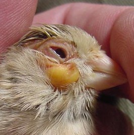
Diagnosis: Frontal Sinus Abscess
Treatment: Mass must be lanced and drained to remove cheesy mass. If undertaken by the bird’s owner, be careful to place pressure on it to stop bleeding. If the bird looses to much blood, it may die. Ideally, this should only be done by an experienced avian technician.
|
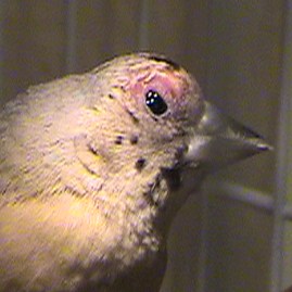 |
Diagnosis: Streptococcus Sinus Infection in juvenile Gouldian
Treatment: Treat for 10 days with Amoxicillin/Tylosin antibiotic. In the blue-back mutation Gouldians these type infections are quite common, especially in the juveniles and is associated with the inherent weakness in this mutation.
|
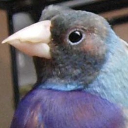 |
Diagnosis: Streptococcus Sinus Infection in Mutation Gouldian.
Treatment: Administer Amoxicillin/Tylosin antibiotic for 10 days. This type infection is quite common in the blue mutation, especially in the juveniles due to an inherent genetic weakness. Stress through the juvenile period will suppress the immune system which precipitates the infection. Usually there will be successive repeat infections and these birds should not be used as breeders.
|
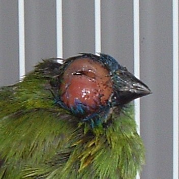 |
Diagnosis: Overwhelming Conjunctivitis with secondary Per orbital Sinus involvement.
Treatment: Treat with an eye cream initially and in water Doxycycline/Tylosin antibiotic mixture for 10 days if no avian vet is available. However, it would be best to have an avian vet culture this to identify the exact cause before any antibiotic is used.
|
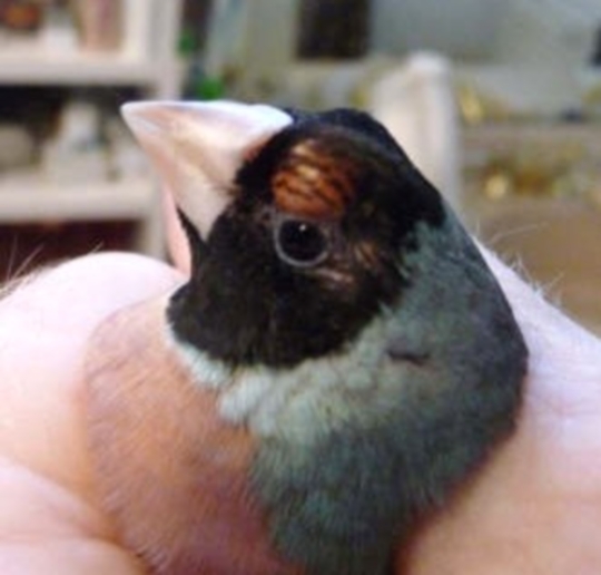 |
Diagnosis: Sinus Infection
Treatment: Swelling must be lanced and the “cheesy abscess” material removed. Pressure must be applied with a cotton bud to stop bleeding. Apply Iodine into wound daily for 3 days. Administer Amoxicillin antibiotic for 7 days. Add vitamin A to the diet as this can be caused by a vitamin A deficiency or a Staphylococcus dust related infection.
|
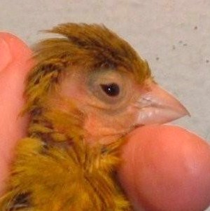 |
Diagnosis: Sinus Infection with possible secondary fungal or Streptococcus skin infection caused by rubbing his face on the perches.
Treatment: Administer Tylosin or Doxycycline/Megamix treatment to clear up the sinus infection. Topical anti-fungal medication if a skin scraping indicates a fungal skin infection. You will need an avian vet to determine the type of skin infection.
|
 |
Diagnosis: Discoloration or darkening of the feathers around the nostrils is caused by a wet discharge from the nostrils.
Treatment: Exact treatment cannot be determined until the reason for the discharge is discovered. I suggest that you see an avian vet to have this bird evaluated.
|
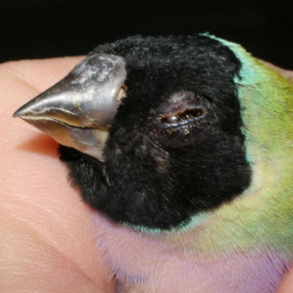 |
Diagnosis: Swollen eye, cause unknown
Treatment: This is one of my hand-fed hens. She was treated for 5 consecutive days with Sovereign Silver drops. I applied the eye drops twice each day for 3 days, then one time each day for 5 additional days. The swelling resolved itself completely.
|
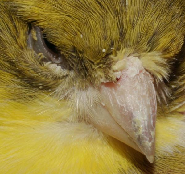 |
Diagnosis: Scaly Face Mites (top of beak and under eye) (Knemidokoptes spp.)
Treatment: Apply undiluted S76 or Scatt using a cotton bud (Q-tip) for one day each week for 6-8 weeks or until the crusty patches fall off. Be careful to avoid the eyes and mouth during application. Scaly Mites attack weak families of birds. The highest quality diet is especially important because Scaly Mites will also attack and infect nutritionally compromised birds.
|
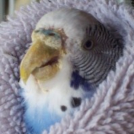 |
Diagnosis: Scaly Face Mites (Knemidokoptes spp.)
Treatment: Apply undiluted S76 or Scatt using a cotton bud (Q-tip) for one day each week for 6-8 weeks or until the crusty patches fall off. Be careful to avoid the eyes and mouth during application. Scaly Mites attack weak families of birds. The highest quality diet is especially important because Scaly Mites will also attack and infect nutritionally compromised birds.
|
 |
Diagnosis: Possibly Scaly Face Mites in advanced stage. (Knemidokoptes spp.)
Treatment: It is impossible to exactly determine the cause of this deformity from the photo. An examination and scraping must be done by an avian vet to determine the exact cause before any treatment is begun.
|
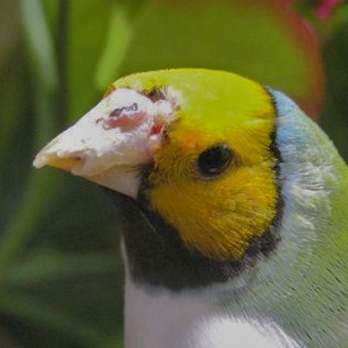 |
Diagnosis: This is another case of being unable to determine the exact cause of this problem.
Treatment: Owner tried treating with an anti-fungal cream with some improvement. It is possible that this is a result of several different infections and would be best handled by an avian vet who could culture the tissues involved.
|
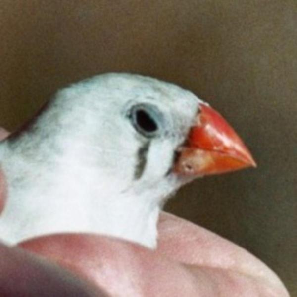 |
Diagnosis: This appears to be a friction granuloma related to a food dish that is too high or in an awkward position.
Treatment: Check possible causes of physical abrasion before looking elsewhere.
|
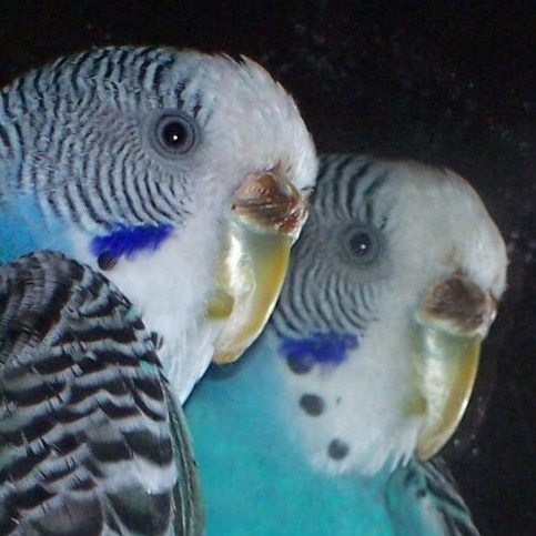 |
Diagnosis: Owner was concerned about color of Cere and overgrown beak
Treatment: Overgrown beak needs regular trimming, but otherwise this Budgie looks to be in good health. Cere color is fine.
|
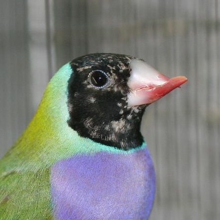 |
Diagnosis: Crossed Beak
Treatment: This is a genetic abnormality which can be passed to this bird’s offspring. Therefore breeding this bird is NOT recommended. With regular trimming of the beak by an owner, this bird will live out a normal life span. White spots in the black head feathers are juvenile feathers since this bird did not successfully complete its juvenile molt. Head should turn completely black after the 1st adult annual molt.
|
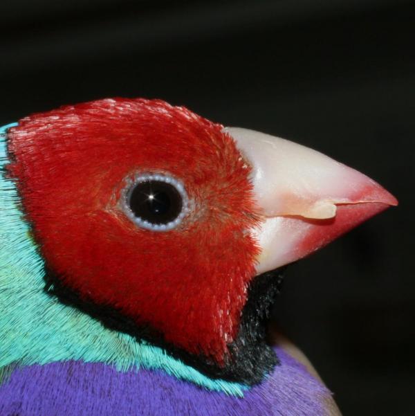 |
Diagnosis: Overgrown “fang” on side of beak
Treatment: I have found this trait to have some genetic factors, but basically it appears to be just a “lazy” bird who does not keep its beak trimmed when rough perches are supplied. This will also happen when the bird has nothing “rough surfaced” in its cage on which to keep the beak trimmed. Owner can trim “fang” with nail clippers, but best to provide appropriate surfaces so bird can trim itself.
|
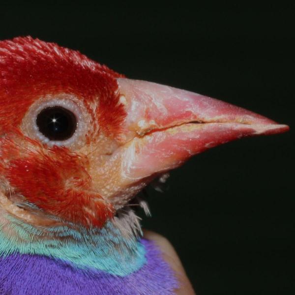 |
Diagnosis: Elongated beak – Polyoma-like virus, NO CURE
Treatment: Exposure to Polyomavirus happened when this bird was a baby. In finches, Polyomavirus is passed only to baby birds by “carrier” adults. However not all carrier birds will show outward signs of Polyomavirus, which makes it hard to determine the adult carrier that is exposing your baby birds. It does not necessarily have to be a parent bird.
|
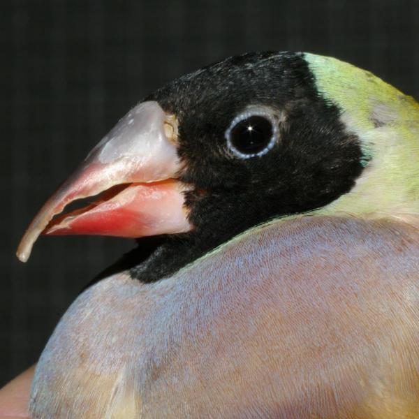 |
Diagnosis: Elongated beak – Polyoma-like virus, NO CURE
Treatment: Exposure to Polyomavirus happened when this bird was a baby. In finches, Polyomavirus is passed only to baby birds by “carrier” adults. However not all carrier birds will show outward signs of Polyomavirus, which makes it hard to determine the adult carrier that is exposing your baby birds. It does not necessarily have to be a parent bird.
|
 |
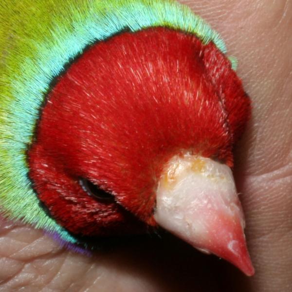
Diagnosis: I do not know the exact cause of these black holes on this bird’s beak but I suspect a mite of some kind
Treatment: You could use either S76 (Ivermectin) or Scatt (Moxidectin) to kill the mites by applying it to the affected area once each day for 1 week, then once each week until the holes disappear. I chose to threat this bird with Organic Neem Oil, everyday for 1 week and then once per week for 5 weeks. (see next photo)
|
 |
Diagnosis:
Treatment: This photo was taken 4 weeks after the one above. Black Holes have closed up but beak was stained by the Neem Oil. I continued treatment with the Organic Neem Oil for 4 more weeks. Stain on beak eventually disappeared as the beak grew out.
|
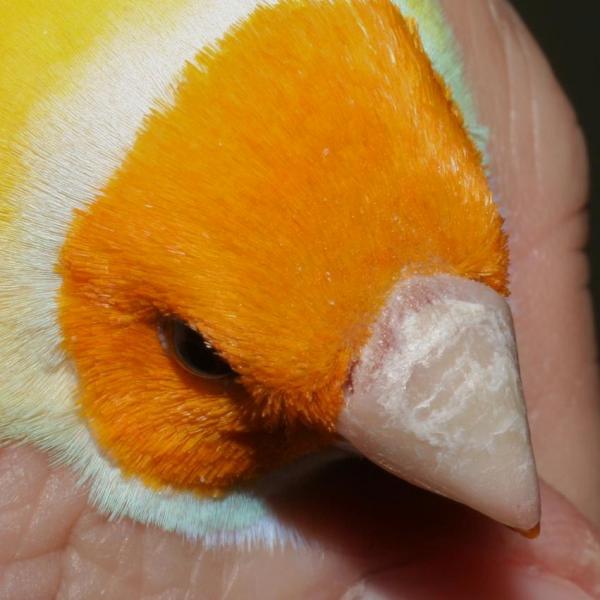 |
Diagnosis: White crusty growth on upper mandible. August 1st
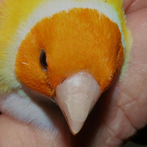
Treatment: This is one of my birds. I treated him with Organic Neem Oil, applied once each day for 7 consecutive days
FOLLOW WITH PICTURE BELOW
|
 |
Diagnosis:
Treatment: This photo was taken September 4th. You can see that the beak is growing out and the crusty patch is no longer showing on the new beak surface (closest to the nostrils).
After the 1st week of treatment with Organic Neem Oil, I applied the oil with a Q-tip one day each week.
|
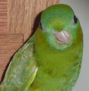 |
Diagnosis: This beak deformity could be congenital or acquired from jaw damage at 7-10 days of age. It can also be associated with sunlight/vitamin D/calcium problems at a young age.
Treatment: No Cure, this change is permanent, but regular beak trimming should allow this bird to eat properly.
|
 |
Diagnosis: Congenital, developmental abnormality, not Polyoma-like virus seen in finches.
Treatment: No treatment possible
|